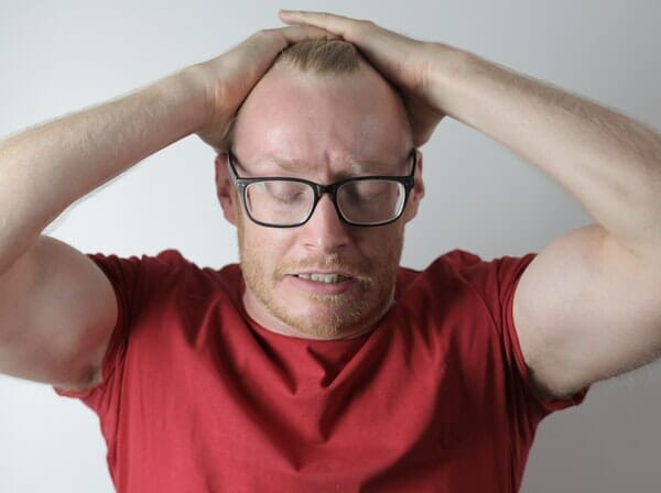
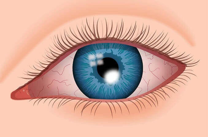
According to the American Society of Cataract and Refractive Surgery (ASCRS), there were approximately 1.8 million LASIK procedures performed in […]
Photophobia or photosensitivity, is one of the more common conditions associated with a traumatic brain injury, experienced by approximately 50% of patients.
One of the most common symptoms reported after a head injury is photophobia or light sensitivity. This is experienced both by those who have had a serious head injury (TBI or traumatic brain injury) as well as a mild head injury (mTBI).
Mild TBI (mTBI), which is typically defined by Glasgow Coma Scale score ≥ 13, accounts for the vast majority of all TBIs, particularly in the setting of sports-related injuries. The Glasgow Coma Score is a method for assessing the patient's extent of consciousness after a traumatic brain injury.
At our specialty eye care center we provide advanced care for patients who experience light sensitivity following a stroke, concussion or neurological condition. We are proud to provide cutting edge neuro optometric rehabilitation services to patients from all over Washington, and we are proud to be a leading provider of neuro-optometric rehabilitation services for patients from Olympia, Lakewood, Tacoma, and Lacey.
A mTBI can result in a variety of visual complications. These are some of the most common visual-related conditions and symptoms:
The conditions described above are in some way related to one another. There is a link between convergence problems and accommodative problems, for example. Another example is headaches and photophobia. Post Traumatic Vision Syndrome was described by William V. Padula, OD, as a cluster of vision and spatial disorders that affect people who have suffered a neurological event, such as a traumatic brain injury, a cerebral vascular accident, multiple sclerosis, or another neurological disorder.
If you are experiencing any of the above symptoms and conditions following a traumatic brain injury, you don't have to suffer any longer. It is possible to alleviate these symptoms with a customized neuro-optometric rehabilitation plan and continue to do your everyday tasks without difficulty and sensitivity to light. To schedule a neuro-optometric evaluation, please call (360) 491-2121.
Often, when the visual system isn't functioning appropriately after a neurological/traumatic injury, we see a high level of exophoria (one eye drifting out) or exotropia (eye misalignment where one eye deviates outwards). Due to this additional effort, the visual system is stressed and activates the proprioceptive fibers in the extraocular muscles, which transmit signals to the trigeminal ganglion indicating the added effort. Nerve signals sent to the trigeminal ganglion activates pain signals through the thalamus and up to the cerebral cortex. Patients may experience headaches, light sensitivity, or dizziness.
Photophobia is closely linked to pain sensation. It is the trigeminal nerve and its nuclei that are responsible for sensations of pain in the head. Several parts of the eye are densely innervated with trigeminal fibers and sensitive to pain, including the conjunctiva, cornea, sclera, and uvea (iris, ciliary body, and choroid). Photophobia is usually caused by painful stimuli to these areas (e.g. corneal abrasion, iritis, uveitis).
On a pathophysiological level, let's examine some of the more common mechanisms. The retina contains photosensitive cells known as rods, cones, and intrinsic photosensitive retinal ganglion cells (ipRGCs). In the human retina, rods and cones are the predominant photosensitive cells. Melanopsin is found in the intrinsic photosensitive retinal ganglion cells. These cells specifically detect luminance levels, but they do not support vision. The ipRGC sends axons to the suprachiasmatic nucleus of the hypothalamus, and these axons control circadian rhythms. In addition, the ipRGC cells also send axons to the Edinger-Westphal nucleus to control the pupil light reflex. In the human retina, there are only 3,000 of these cells, representing .2% of all RGCs. These cells are photoreceptive, with maximum sensitivity to blue light within the 460-480 nm range. A patient with a traumatic brain injury may have a poorly regulated melanopsin-signaling system when exposed to light.
In patients with photosensitivity, visual fields are important. It was noted by Christine T. Langerhorst and Avinoam B. Safran that there were generalized constrictions in the visual field. While the authors attributed it to an attention disorder, Jackowski attributed it to loss of rod-mediated activity related to dark adaptation. All patients reporting photosensitivity had visual field losses within the central 30 degrees when Jackowski used frequency doubling technology (FDT). Rods affect magnocellular loss, which affects the FDT. These patients reported collapsing visual fields during testing. Kinetic fields, such as Octopus or Goldmann, can also be used to map the visual field constriction. It was found that the FDT and kinetic fields show a strong correlation with visual field constriction.
The degree of light sensitivity caused by mTBI varies from mild to severe. The most severe cases might require patients to wear sunglasses constantly, even indoors. They may be sensitive to fluorescent lighting as well as digital devices, which emit a lot of blue light. With the introduction of filters and vision rehabilitation, light sensitivity can be improved even when it is severe and of long duration.
The goal is to relieve symptoms with specific filters and to take the patient off the use of sunglasses indoors. Photosensitivity is associated with visual fatigue. It often occurs in conjunction with binocular vision problems, and is not a standalone condition. Binasal occlusion is used to reduce binocular stress in patients with mTBI. Through binasals, the system becomes less hyper-focused, which reduces sensitivity to light. Additionally, blue filters may reduce symptoms of indoor light sensitivity. Polaroid, blue-blocking filters, and FL-41 filters can be used outdoors to tone down the brightness.
Photophobia is a complex subject. Research in the future will provide us with a deeper understanding of photophobia in mTBI patients and allow us to provide better treatment for them.
If you or a loved one has suffered a traumatic brain injury and has been suffering from light sensitivity or any of the above listed visual conditions, please schedule a neuro-optometric evaluation with Dr. Zurcher.
Originally from Arizona, Levi received his Bachelor’s of Science in Zoology from Northern Arizona University and his Doctorate in Optometry from Pacific University in Oregon. He is board certified in developmental vision and vision therapy by COVD. He purchased the practice from the Inversos in 2012. He likes spending time with his family (wife and two children,) mountain biking, and undertaking creative projects. He wrote the vision therapy software used by the clinic, retinal camera software, and the computerized eye-chart used for exams.
To schedule a neuro optometric evaluation, please call (360) 491-2121. Those who have suffered a traumatic brain injury and have been suffering from visual conditions visit our clinic from all over Washington, and we are proud to be a leading provider of neuro-optometric rehabilitation services for patients from Olympia, Lakewood, Tacoma, and Lacey.
Such a nice optician. My grandson is only 4 and needs glasses. We were so sad, but he explained the issues, and we will follow up as he suggested.
I had such a good experience with the clinic. Very friendly staff and doctor, did not have to wait for long to be called, and was treated respectfully. Thanks, American Family.
Dr. Zurcher has gone above and beyond what any other eye doctor has ever done to figure out what is going on with my eyes. Very happy with American Family Vision.
Family Vision Clinic changed our lives! My daughter was frequently car sick, and she was getting headaches every day, often painful enough that they brought her to tears. We saw a string of doctors and therapists, but we made no progress. Finally, we found Dr. Levi Zurcher and his eye therapist Rain. After ten weeks of eye therapy my daughter no longer gets headaches, and she no longer gets car sick. Daily tears are a thing of the past. I really can’t say enough about this clinic. It was fascinating to watch Dr. Zurcher work. For the first time, someone who knew what they were doing was intently studying my daughter, really trying to figure out all of her eye issues, and his therapist Rain is one of the most patient and lovely people that I’ve ever had the good fortune to meet. We’re finished with eye therapy—yay!—and I have switched to American Family Vision Clinic for all of my family’s other eye health needs.
Very professional, yet kind and helpful. They do what they can to make the appointment comfortable. I was running a bit late, I made sure ti call. They were able to switch me with a patient who was already there, they treated me with respect, and helped my son have confidence by getting him the eye care he needed that day. Thank you so much! Would recommend to anyone. It's a blessing that they care enough to work with people who have all different types of insurance from work to state coverage.
The staff is friendly. The Doc is very knowledgeable. The office is family friendly and everyone is so patient with the little ones.
According to the American Society of Cataract and Refractive Surgery (ASCRS), there were approximately 1.8 million LASIK procedures performed in […]
As a local optometry clinic, we often get asked how to get rid of under eye bags. Many people have […]
Bacterial conjunctivitis is a prevalent condition globally. In the US, it affects 135 out of every 10,000 people each year, […]
Monday-Thursday
8:30AM-5:00PM
Friday
8:30AM-3:00PM
Saturday-Sunday
Closed